In the realm of medical diagnostics, precision and speed are paramount, and a recent breakthrough in endomicroscopy technology is set to redefine the standards for gastrointestinal inspections. Researchers, led by Jintaek Im from the Department of Robotics and Mechatronics Engineering at DGIST in South Korea, have developed a compact, multimodal endomicroscope that combines fluorescence (FL) and optical coherence tomography (OCT) imaging into a single, forward-viewing probe. This innovation, detailed in the *International Journal of Optomechatronics* (which translates to the Journal of Light and Machine Integration), promises to enhance diagnostic accuracy and expedite decision-making processes in clinical settings.
The demand for real-time, precise diagnostics has been growing, particularly in the field of gastrointestinal endoscopy. Current endoscopy techniques, while effective for wide-area inspections, often fall short in providing detailed tissue morphology and tumor invasion depth. Traditional histological analysis through biopsy, though comprehensive, is time-consuming and can delay critical treatment decisions. Im and his team recognized this gap and set out to bridge it.
“The integration of FL and OCT modalities within a single probe allows for a more comprehensive and immediate assessment of tissue morphology and structure,” Im explained. “This dual approach not only enhances diagnostic accuracy but also supports real-time decision-making, which is crucial in clinical settings.”
The newly developed endomicroscope employs a Lissajous scanning mechanism to achieve uniform illumination and high-speed imaging. The probe itself is a marvel of compact design, assembled from a composite fiber, a piezoelectric tube actuator, and an asymmetrically attached polymer stiffener. This configuration enables combined fluorescence and optical coherence imaging with complementary performance characteristics.
One of the standout features of this technology is its compatibility with standard gastrointestinal endoscopic channels. This means it can be seamlessly integrated into existing medical practices without the need for extensive modifications. The system also incorporates parallel computing for real-time image reconstruction, ensuring high-throughput data processing.
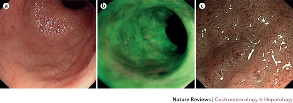
Imaging experiments conducted on phantom targets and ex-vivo animal tissues have confirmed the system’s capability to produce detailed, co-registered images of tissue morphology and structure. These results underscore the potential of the technology to revolutionize diagnostic procedures in gastrointestinal endoscopy.
The implications of this research extend beyond the medical field. In the energy sector, for instance, similar imaging technologies could be adapted for inspecting and maintaining complex infrastructure, such as pipelines and turbines. The ability to perform real-time, high-resolution imaging could significantly enhance safety and efficiency in these applications.
As the technology continues to evolve, it is poised to shape the future of diagnostic and inspection procedures across various industries. The work of Jintaek Im and his team, published in the *International Journal of Optomechatronics*, represents a significant step forward in the field of optomechatronics, offering a promising platform for enhancing diagnostic accuracy and enabling real-time decision-making.
In an era where precision and speed are paramount, this innovation stands as a testament to the power of interdisciplinary research and its potential to transform industries. As we look to the future, the integration of advanced imaging technologies like this multimodal endomicroscope will undoubtedly play a pivotal role in shaping the landscape of diagnostics and beyond.