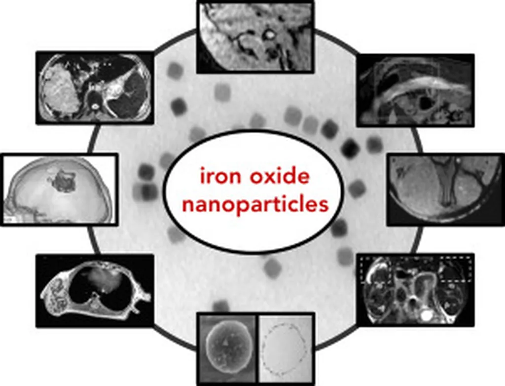
In the ever-evolving landscape of medical imaging, a groundbreaking study led by Aleia G. Williams from the Department of Biomedical Engineering at the University of Tennessee Space Institute has shed new light on the potential of magnetic particle imaging (MPI). This innovative technique, which uses magnetic nanoparticles as tracers, could revolutionize the way we detect and monitor various medical conditions, with significant implications for the energy sector as well.
MPI offers a unique advantage over traditional magnetic resonance imaging (MRI) by directly detecting the magnetization of nanoparticles rather than relying on secondary effects. “This direct detection method can lead to more precise and faster imaging,” Williams explains. However, the size of the nanoparticles used in MPI has been a limiting factor in achieving optimal performance.
Williams and her team hypothesized that pure iron nanoparticles could enhance MPI performance, but creating larger iron nanoparticles has proven challenging. Previous studies have typically produced nanoparticles in the 5-15 nm range. To overcome this hurdle, the researchers turned to an extended LaMer mechanism, a process that involves the continuous addition of a precursor to control particle growth.
The team tested three different injection speeds of the iron precursor, Fe(CO)5, and three different surfactants to determine their effects on nanoparticle size, morphology, and composition. The results were promising. “We observed larger nanoparticle diameters up to 24 and 26 nm at lower injection speeds when using hexadecylamine (HDA) and octadecylamine (ODA) surfactants, respectively,” Williams notes. However, samples using an oleylamine/oleic acid (OAm/OA) mixture remained around 15.5 nm at all injection speeds.
The study, published in the journal *Academia Materials Science* (translated to English as “Academic Materials Science”), also revealed that while OAm/OA samples displayed high magnetic saturation values, samples using HDA and ODA showed lower magnetic saturation values. This trade-off between size and magnetic saturation presents an interesting dilemma for researchers aiming to optimize MPI tracers.
The implications of this research extend beyond the medical field. In the energy sector, MPI could potentially be used for real-time monitoring of pipelines and other infrastructure, detecting leaks or corrosion before they become critical. The development of larger, more effective nanoparticles could enhance the sensitivity and accuracy of these applications.
As Williams and her team continue to explore the potential of iron nanoparticles in MPI, their work could pave the way for advancements in both medical imaging and industrial monitoring. The journey towards optimizing MPI tracers is ongoing, but with each step, we move closer to unlocking the full potential of this groundbreaking technology.